Description
Financing Available
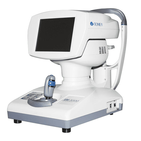
Tomey EM 3000 Specular Microscope
Serial photographs of 15 shots
15 shots can be taken in series and errors during photographing are reduced. In addition, the best image among the shots is automatically selected and displayed on the screen.
Wide photographing range and 7 capturing positions
Our unique technology enables a wide photographing range of 0.25 x 0.54 mm and allows you to observe the endothelium over a wide range.
Photos can be taken at 7 points: the center and 6 peripheral points (2, 4, 6, 8, 10, and 12-o’clock positions on a 0 6 mm arc). Because there are many photographing points, you can select the point with the best conditions even when the cornea surface is irregular. The cornea thickness is also measured at the same time.
Quick and automatic analysis of corneal endothelium cells
The software for automatic analysis is pre-installed, so images are analyzed automatically without personal computers.
Manual photographing is also available
When automatic photographing is difficult, you take photos manually using the power joystick.
LED light source
A long-life LED has been introduced for the photographing light source instead of the conventional xenon lamp, which requires maintenance. Regular replacement of the lamp is a thing of the past.
USB connector for printer and LAN connector for PC
USB-D connector: Connected to a Pict Bridge compatible printer to print images of the corneal endothelium and analysis results
USB-H connector: Connected to a barcode reader or electromagnetic card reader to enter patient ID data. A digital printer may also be connected
LAN connector: After installing the “Data Transfer” software provided with the
EM-3000 in your personal computer, inspection result files assigned a patient ID can be saved in the personal computer.
