Used Ophthalmic Diagnostic Equipment
As an eye care professional, you require a bevy of sophisticated and expensive ophthalmic diagnostic equipment in order to achieve the best possible visual outcomes for your patients. In addition to standard slit lamps, phoropters, tonometers and autorefractors, there are more options to consider now than ever before. Many technologically advanced devices that were introduced in recent years have dramatically changed how physicians diagnose and treat eye diseases such as glaucoma, macular degeneration and diabetic retinopathy.
Keeping pace with the latest ophthalmic advances can greatly affect the level of care you provide, as well as your budget. Therefore, the process of finding the ophthalmic diagnostic equipment that best meets the needs of your practice can be daunting. Turn to Vision Equipment Inc. Founded by Leo Hadley, Jr., a respected eye care industry veteran of over three decades, our company is your trusted source for used, professionally refurbished ophthalmic diagnostic equipment.
What We Offer
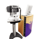
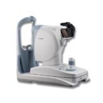
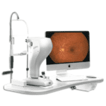
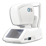
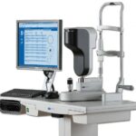
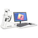
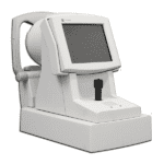
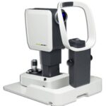
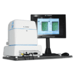
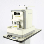
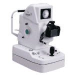
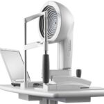
Our expansive inventory of used ophthalmic diagnostic equipment includes:
- A-scan biometers
- B-scan biometers
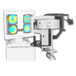
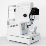
- Corneal topography systems
- Fundus cameras (mydriatic)
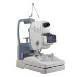
- Fundus cameras (non-mydriatic)
- Ophthalmoscopes
- Pachymeters
- Perimeters
- Refractors
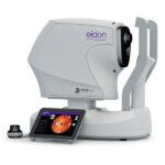
- Retinal imaging devices
- Slit lamps
- Specular microscopes
- Optical coherence tomographers
- Tonometers
Save on Your Equipment Purchases
Many new ophthalmic diagnostic devices cost tens of thousands of dollars. But, you can utilize certain strategies to save money on your equipment purchases. For instance, our used, professionally refurbished ophthalmic diagnostic equipment is available at a significant discount. With us, you also have the option of trading in an older device for credit toward the purchase of a newer model. Finally, we offer attractive financing options to make it easier for you to purchase the ophthalmic diagnostic equipment you need for your practice.
Technological advances are occurring very rapidly in the field of ophthalmology, which can make it especially difficult to know when it is best to invest in a product or wait for a possible new version on the horizon. What’s more, there is a general lack of associations that fully understand the complex needs of today’s ophthalmologists. At Vision Equipment Inc., our business is exclusively dedicated to buying, selling, trading and refurbishing high-quality ophthalmic equipment. We can help you build your practice and become a trusted partner in your success.
To learn more, contact Vision Equipment Inc. today.
Showing 1–20 of 101 results