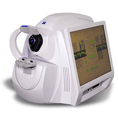
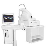
ZEISS CIRRUS 500 HD-OCT
Financing Available

Used ZEISS CIRRUS 500 HD-OCT
A robust optical coherence tomography (OCT) machine, the ZEISS CIRRUS 500 HD-OCT features top-of-the-line visualization, tracking and assessment tools for a broad range of clinical applications, including glaucoma management, retinal assessment for cataract surgery and anterior segment imaging for corneal disease. By purchasing a used OCT from Vision Equipment Inc., you can optimize your eye care practice with industry-defining technology while saving thousands of dollars.
Highly Accurate Imaging
As compared to its predecessors, the ZEISS CIRRUS 500 HD-OCT is a much faster OCT scanner. As a result, this easy-to-operate instrument makes it much simpler to properly align a patient for highly accurate imaging.
Revolutionary Assessment Options
The ZEISS CIRRUS 500 HD-OCT allows you to compare up to six progression maps simultaneously. For ease of analysis, areas of statistically significant change are color-coded, enhancing your ability to collect macular, optical nerve head (ONH) and retinal nerve fiber layer (RNFL) information and track changes over time.
Multi-Angle Visualization
Thanks to revolutionary three-dimensional imaging, advanced visualization and OCT fundus images, the ZEISS CIRRUS 500 HD-OCT allows you to view cube data from multiple angles. Focusing on millions of data points and tightly spaced B-scans, this sophisticated OTC machine can image even the smallest areas of pathology.
Buy Used & Save
The ZEISS CIRRUS 500 HD-OCT 500 is an outstanding solution for an ophthalmic practice. If you’re in the market for one, you can buy used – with complete confidence – from Vision Equipment Inc. Our factory-trained technicians meticulously return used ophthalmology equipment to like-new condition. Contact us today.
Description
ZEISS CIRRUS 500 HD-OCT
ZEISS CIRRUS HD-OCT 500 provides a great solution for comprehensive ophthalmic practices and offers essential OCT capabilities with a broad range of clinical applications in an easy-to-learn, easy-to-use instrument for the management of glaucoma and retinal disease, retina assessment for cataract surgery, and anterior segment imaging for corneal disease. The new OCT camera enables quick OCT fundus image refresh making patient alignment more efficient.
Advanced diagnostic tools that improve your ability to identify pathology and track change over time.
- NEW Macular Thickness OU Analysis
- Ganglion Cell Analysis
- Guided Progression Analysis (GPA™)
- Macular Thickness and Change Analysis
- Macular Thickness Normative Data
- 34 kg
- For Iris Imaging : 1280 x 1024
- (WxDxH) 18 x 26 x 21 in. Table: 39L x 22W
- Internal, external
- -20D to +20D (diopters)
