Description
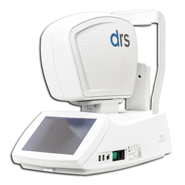
DRS Non Mydriatic Fundus Camera
Thanks to its fully automated operation, the DRS fundus camera requires minimal operator training. Its compact, ergonomic design and low power flash help ensure patient comfort. DRS is conceived to maximize patients flow work and it is entirely operated through its intuitive touch-screen interface. It supports single -or multi- field acquisition protocols, providing seven different 45° fields. It can be used both for screening and diagnosis.
Benefits of DRS:
- Extreme ease of use: patient auto-sensing, auto-alignment, auto-focus, auto flash adjustment, auto-capture
- Short exam time: captures both eyes in 1 min (single field)
- High quality images
- Minimal training required
- Compact & clean design
- No additional PC required
- Touch-screen operation
- View images from any PC or tablet
- Internet-connected
- Wi-Fi and Ethernet connectivity
- Export to USB and remote folder (for import into EMR), DICOM, JPG, PDF
| Retinal Imaging | |
| Field of view | 45° x 40° |
| Non mydriatic operation | (4.0 mm minimum pupil size) |
| Fixation target | 7 internal LEDs |
| Operating distance | 37 mm |
| Sensor size | 5 MPixel (2592×1944) |
| Sensor resolution | 48 pixels/deg |
| Dimensions | |
| Weight | 19 Kg (42 lbs) |
| Size | 58 x 55 x 33 cm (23’ x 22’ x 13’) |
| Other Features | |
| Power cord, spare fuses, dust cover | |
| Patient presence sensor | |
| Motorized chin-rest | |
| Automatic alignment using two pupil cameras | |
| Auto-focus (adjustment range -15D to +15D) | |
| Auto-flash level adjustment | |
| Low power flash | |
| 10.4” touch-screen color display | |
| Embedded PC (160 GB hard disk) | |
| Wi-Fi and Ethernet connectivity | |
| Export to remote folder (for import into EMR) | |
| Multiple fields acquisition | |
| Stereo Pair | |
| External Eye |
